
Hello, this is Goyang Shinsegae Eye Clinic.
Many people dismiss slightly blurred or hazy vision as a temporary sign of fatigue.
When vision declines with age, it is also easy to assume that it is simply a natural part of aging.
However, diabetic retinopathy, macular degeneration, and glaucoma often cause few noticeable symptoms in their early stages.
If you experience blurred vision, distorted images, floating spots or strands, or a narrowing of your visual field, it is advisable to visit an eye clinic in Haengsin-dong for an examination and consultation.
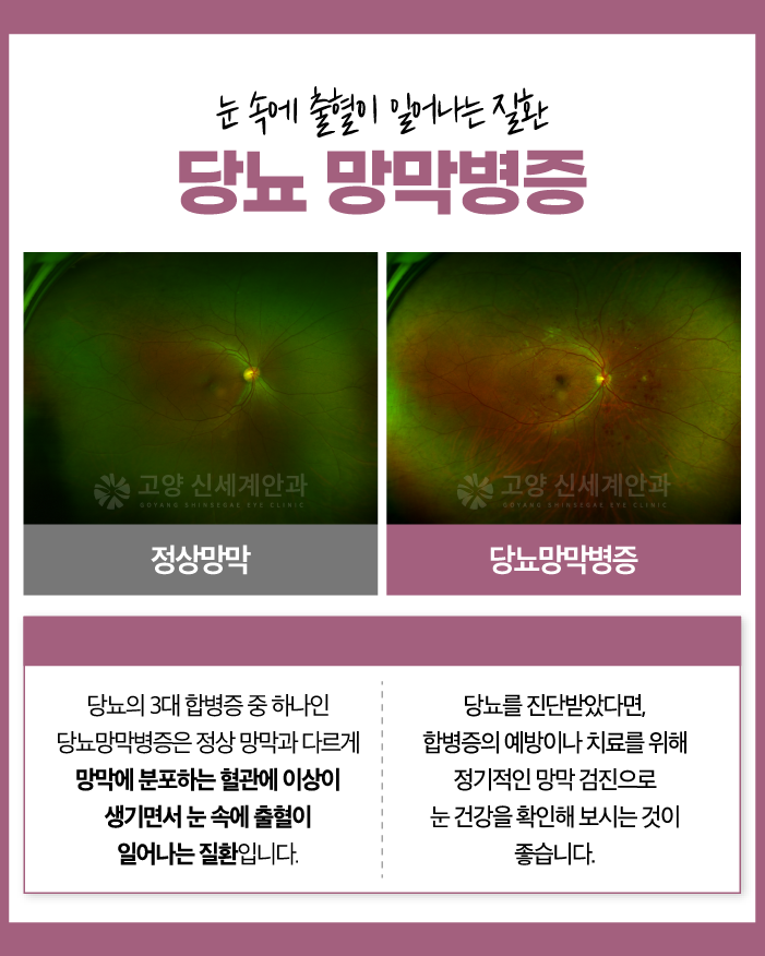
Type 01. Diabetic Retinopathy
Diabetic retinopathy is one of the major microvascular complications associated with diabetes.
It can develop when persistently high blood glucose levels damage the small blood vessels in the retina.
The retina is an essential layer of neural tissue that receives light entering the eye and transmits visual information to the brain.
When blood glucose remains poorly controlled, the walls of the retinal blood vessels may weaken, allowing blood or fluid to leak.
As the condition progresses, retinal bleeding or swelling may occur. A lack of oxygen in the retina may also lead to the formation of abnormal new blood vessels.
This process can cause hazy vision, floaters that appear as dots or threads, and a sudden decline in vision.
Central vision may also deteriorate if macular edema develops.
What Are the Treatment Options?
✅ Focal and Panretinal Photocoagulation
This treatment uses a laser to create controlled burns in selected areas of the retina.
It may be used to reduce the risk of further bleeding and suppress the growth of abnormal blood vessels.
✅ Intravitreal Injections
Anti-vascular endothelial growth factor, or anti-VEGF, medication is injected into the vitreous cavity inside the eye.
This treatment can inhibit the growth of abnormal new blood vessels and help reduce macular edema.
✅ Vitrectomy
Surgical treatment may be required when severe vitreous hemorrhage or tractional retinal detachment develops.
A vitrectomy removes blood and cloudy material from the vitreous cavity to help stabilize the retina.

Type 02. Macular Degeneration
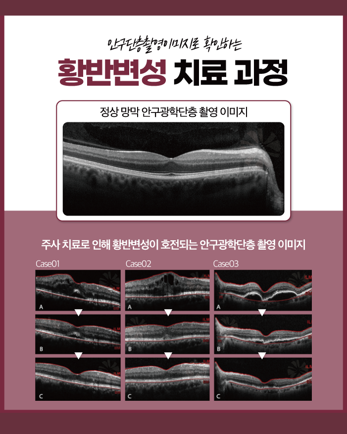
The macula is located at the center of the retina. It plays an important role in reading, seeing objects clearly, and distinguishing colors.
Macular degeneration is a condition in which changes in the macula lead to a decline in central vision.
The likelihood of developing the condition increases with age. People with risk factors such as a family history of macular degeneration or smoking should be particularly cautious.
One of the most common symptoms is metamorphopsia, in which straight lines or grid patterns appear bent or distorted.
The center of the visual field may also appear blurred, dark, or partially blocked.
What Are the Treatment Options?
✅ Photodynamic Therapy
Photodynamic therapy, or PDT, involves administering a photosensitizing medication and applying a specific type of laser to the affected area.
✅ Laser Photocoagulation
A laser is used to seal abnormal new blood vessels.
✅ Intravitreal Injections
Anti-VEGF medication is injected into the eye to suppress the growth of abnormal blood vessels and reduce retinal bleeding or swelling.
✅ Vitrectomy
Surgical treatment may be considered when extensive bleeding occurs or blood accumulates in the vitreous cavity and affects vision.
Type 03. Glaucoma
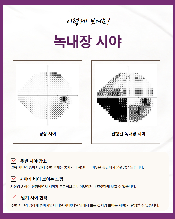
Glaucoma is a condition in which progressive damage to the optic nerve leads to a gradual narrowing of the visual field.
If the damage becomes advanced, central vision may also be affected. Early detection and consistent management are therefore important.
Glaucoma is often associated with elevated intraocular pressure. However, normal-tension glaucoma can develop even when eye pressure remains within the statistically normal range.
In addition to intraocular pressure, several other factors may contribute to glaucoma, including structural vulnerability of the optic nerve, impaired blood flow, myopia, and family history.
Most importantly, glaucoma often causes no obvious symptoms in its early stages.
Because damage may begin gradually in the peripheral visual field, a comprehensive assessment of the optic nerve is necessary.
This may include fundus examination, optical coherence tomography (OCT), visual field testing, and ganglion cell layer analysis.

What Are the Treatment Options?
✅ Medication
Pressure-lowering eye drops are used to reduce intraocular pressure and slow further damage to the optic nerve.
✅ Trabeculectomy
Trabeculectomy may be considered when medication or laser treatment does not lower intraocular pressure sufficiently.
This procedure creates a new drainage pathway for aqueous humor to leave the eye, thereby lowering intraocular pressure.
✅ Laser Peripheral Iridotomy
A laser is used to create a small opening in the iris. This improves the flow of aqueous humor and helps regulate intraocular pressure.

If you have diabetes, a family history of glaucoma or macular degeneration, high myopia, or changes in your vision that differ from what you previously experienced, regular examinations at an eye clinic in Haengsin-dong are recommended.
Periodic eye examinations can help detect changes earlier and allow appropriate treatment to begin at the right time.